Monday, November 22, 2010
Microscopes
Last week was really fun. We got to use the microscopes. I got really good at focusing the microscope in on what we are looking at. I fought for the microscope camara. So we got some really good pictures of onion cells and cheek cells. we tried to blow up the cells by putting distilled water and shrink the cells using salt water. The cells grew and shrunk a little bit but not by much. The first day we were using them we looked at pond water and you could see little microorganisms moving around. Also I was bleeding so i put a little of my blood on a slide and we looked at that. It was cool you can see all the blood cells. I can't wait to see what we get to look at next through the microscopes. Check out the following video from the microscope camera.

Friday, November 19, 2010
CYSTIC FIBROSIS
CYSTIC FIBROSIS - A DISORDER OF MEMBRANE TRANSPORT
A great variety of proteins have roles in moving molecules and ions across cell membranes. Passive transport proteins permit certain substances to diffuse down concentration gradients by moving through the protein's interior. Active transport proteins use ATP energy to pump substances across the membrane against their concentration gradients. To investigate the importance of transport proteins, we will consider the effects of cystic fibrosis, a genetic disorder in which there is a defect in a transport protein..
Activity
Part 1.
In this part of the activity you will visit the Cystic Fibrosis Foundation’s web site to learn about the causes and symptoms of cystic fibrosis.
Use your browser to go to http://www.cff.org/home/
Use the information provided in the “About cystic fibrosis” section to answer the following questions:
1. What are the signs and symptoms of cystic fibrosis?
The symptons of cf of are salty tasting skin, caughing at time with plegm, frequent lung infections, shortness of breath, poor growth/weight gain in spite of good appitite, frequent greasy bulky stools or difficult bowel movements.
2. How common is this disorder?
About 1000 new causes are diagnossed each year.
3. How is cystic fibrosis diagnosed?
A sweat test can be used to diagnose a patient with cf. a high level of salt indicated cf.
4. How is cystic fibrosis inherited? Does everyone who has a mutant gene for the protein have cystic fibrosis?
Cf is inheretied through a mutant protein gene. No there is a 25% chance that they will not inherat cf.
Part 2.
In this part of the activity you will read an article to learn more about cystic fibrosis.
Use your browser to go to:
http://resources.schoolscience.co.uk/MRC/3/page3.html
Use the information in this article to answer the following questions:
1. Explain the normal function of the protein that is defective in cystic fibrosis.
There are channels in these lining cells through which ions can pass. Normally, the movements of ions brings water to the surface of the airway and keeps the mucus moist.
2. What happens to this protein in CF patients and what are the consequences for the health of these individuals?
The defective gene acts to block the channels, which causes the mucus to dry out. It is then difficult for a person to shift the mucus, which then becomes prone to infection by bacteria.
Part 3.
In this part of the activity you will read about how cystic fibrosis is treated.
Use your browser to go to:
http://www.mayoclinic.com/health/cystic-fibrosis/DS00287
Use the information in the “Treatment” and “Self-care” sections to answer the following questions:
1. Explain at least 3 treatments for the symptoms of cystic fibrosis.
One treatment they can give cf patients in medication. there are antibotics, mucus thinning drugs and Bronchodilators. they help stop lung infection, thins the mucus and makes it less sticky, and keeps the airway open.
Another treatment is Therapy. it physically loosens the mucus in the chest.
The last treatment is Surgical. like feeding tubes lung transplants and bowel surgery.
2. Discuss at least 3 ways for parents to help their children who have cystic fibrosis.
Parents should talk openly about there feelings. also the should join a support group for the child and last they should get there kid up and moving get them to loosen the mucus in there chest.
A great variety of proteins have roles in moving molecules and ions across cell membranes. Passive transport proteins permit certain substances to diffuse down concentration gradients by moving through the protein's interior. Active transport proteins use ATP energy to pump substances across the membrane against their concentration gradients. To investigate the importance of transport proteins, we will consider the effects of cystic fibrosis, a genetic disorder in which there is a defect in a transport protein..
Activity
Part 1.
In this part of the activity you will visit the Cystic Fibrosis Foundation’s web site to learn about the causes and symptoms of cystic fibrosis.
Use your browser to go to http://www.cff.org/home/
Use the information provided in the “About cystic fibrosis” section to answer the following questions:
1. What are the signs and symptoms of cystic fibrosis?
The symptons of cf of are salty tasting skin, caughing at time with plegm, frequent lung infections, shortness of breath, poor growth/weight gain in spite of good appitite, frequent greasy bulky stools or difficult bowel movements.
2. How common is this disorder?
About 1000 new causes are diagnossed each year.
3. How is cystic fibrosis diagnosed?
A sweat test can be used to diagnose a patient with cf. a high level of salt indicated cf.
4. How is cystic fibrosis inherited? Does everyone who has a mutant gene for the protein have cystic fibrosis?
Cf is inheretied through a mutant protein gene. No there is a 25% chance that they will not inherat cf.
Part 2.
In this part of the activity you will read an article to learn more about cystic fibrosis.
Use your browser to go to:
http://resources.schoolscience.co.uk/MRC/3/page3.html
Use the information in this article to answer the following questions:
1. Explain the normal function of the protein that is defective in cystic fibrosis.
There are channels in these lining cells through which ions can pass. Normally, the movements of ions brings water to the surface of the airway and keeps the mucus moist.
2. What happens to this protein in CF patients and what are the consequences for the health of these individuals?
The defective gene acts to block the channels, which causes the mucus to dry out. It is then difficult for a person to shift the mucus, which then becomes prone to infection by bacteria.
Part 3.
In this part of the activity you will read about how cystic fibrosis is treated.
Use your browser to go to:
http://www.mayoclinic.com/health/cystic-fibrosis/DS00287
Use the information in the “Treatment” and “Self-care” sections to answer the following questions:
1. Explain at least 3 treatments for the symptoms of cystic fibrosis.
One treatment they can give cf patients in medication. there are antibotics, mucus thinning drugs and Bronchodilators. they help stop lung infection, thins the mucus and makes it less sticky, and keeps the airway open.
Another treatment is Therapy. it physically loosens the mucus in the chest.
The last treatment is Surgical. like feeding tubes lung transplants and bowel surgery.
2. Discuss at least 3 ways for parents to help their children who have cystic fibrosis.
Parents should talk openly about there feelings. also the should join a support group for the child and last they should get there kid up and moving get them to loosen the mucus in there chest.
Wednesday, November 17, 2010
Membrane structure poster

This is a picture my group drew of the cell membrane. The cell membrane is made up of phospolipids because they are hydrophobic. Hydrophobic means water fearing. This makes them a good barrier for the inside of the cell. Our model shows three different types of protein, the receptor protein, the transport protein and the recognition protein. The receptor proteins receive information from other cells. The transport protein transports allows molecules to come inside the cell. The recognition protein recognizes lets other cells know what it is and what animal it is from.
Monday, October 25, 2010
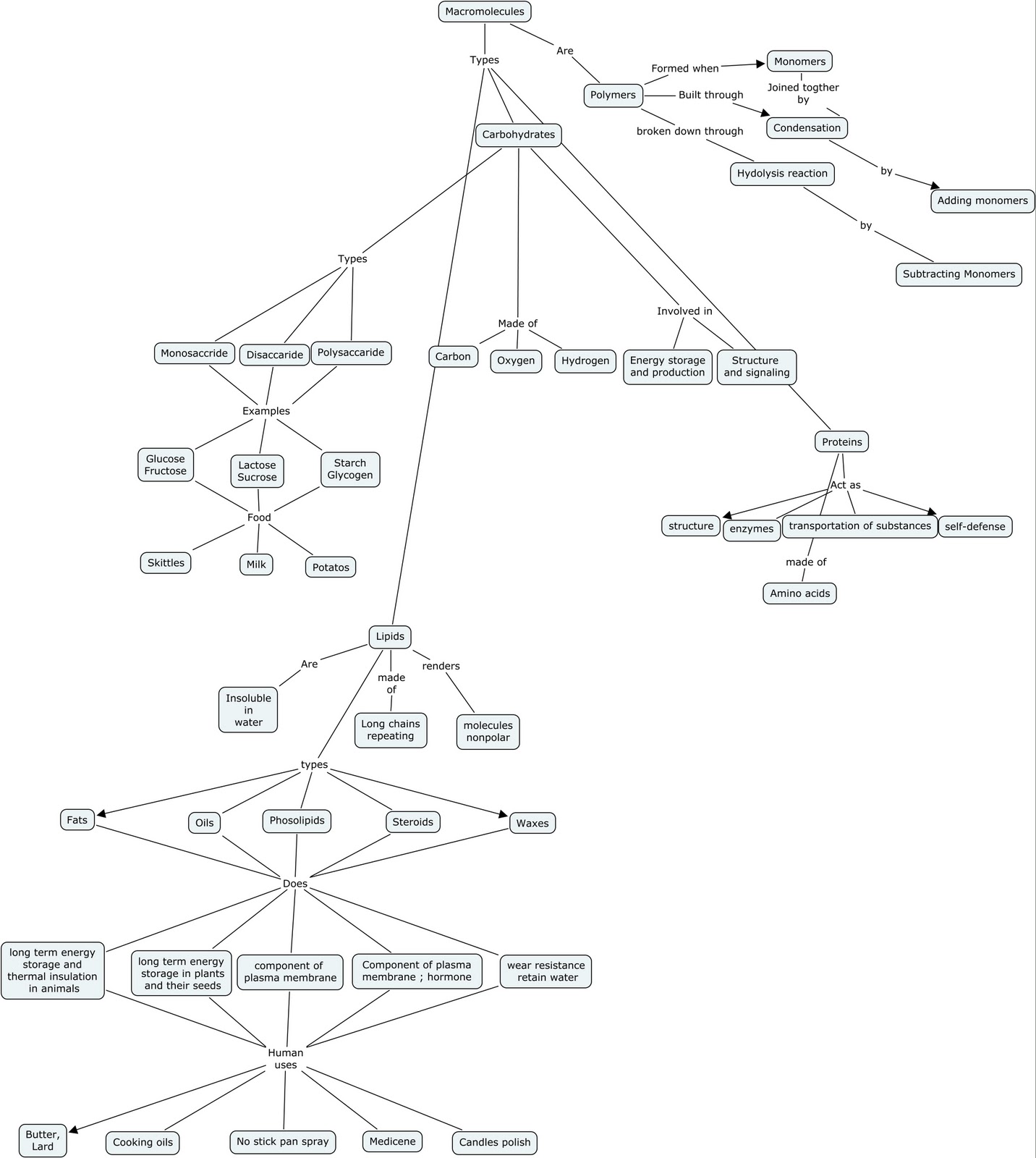
Wednesday, October 20, 2010
Collagen
COLLAGEN STRUCTURE AND FUNCTION
Proteins are the most diverse class of biological molecules. Most of your body is made up of proteins. Each protein consists of one or more polypeptide chains, in which amino acids are strung together in a specific order. This amino acid sequence or 'primary structure' is the key to the shape and function of the protein. Interactions among different amino acids along the length of the polypeptide chain cause it to coil, bend, and fold into a complex shape. Some proteins consist of two or more polypeptide chains. In many cases, the polypeptide chains form a rounded or globular shape. Most enzymes are like this, as is the oxygen-carrying hemoglobin molecule. In other cases, polypeptide chains form long strands or sheets. These fibrous proteins are often structural components, such as keratin and collagen.
Activity
In this activity, you will explore the structure and function of collagen, the most abundant protein in the human body. You will begin by visiting a Protein Data Bank site, where the structure of collagen is described and illustrated in a "Molecule of the Month" feature. Next, you will research the effects of osteogenesis imperfecta (OI), a genetic disorder that affects the structure or number of collagen molecules.
Part 1.
Use your browser to go to
Find the link in the middle of the page to “previous features” under the molecule of the month section. Click there to find the article on collagen in the alphabeticall listing of molecules.
Read the information and view the graphics. Use this information to answer the following questions:
1. Describe the primary structure of collagen. What are the major amino acid components?
The primary action is to strengthen the tendons and vast, resilient sheets that support the skin and internal organs. Every third acid is glycine.
2. What role does vitamin C play in collagen formation? What happens when a person does not get enough vitamin C in his or her diet?
Hydroxyproline is made. If a person doesn’t have enough vitamin c they devolop scurvy
3. Describe the quaternary structure of collagen (the way in which the polypeptide chains are arranged).
Collegen is arranged in a triple helix. Every third amino acid is glycine
Part 2.
Use your browser to go to the Web site of the Osteogenesis Imperfecta Foundation at
On the left side of the home page, click the link “About OI”, then find the “Facts About OI” section.
Read the material in the sections entitled "Facts on Osteogenesis Imperfecta” and “Types of OI.” Use what you have learned to answer the following questions:
1. What is the main symptom of osteogenesis imperfecta? What are some other symptoms that people with OI may have?
The main sympton is fragile bones. They also have muscle weakness, hearing loss, fatigue, joint laxity, curved bones, scoliosis, blue sclerae, dentinogenesis imperfecta (brittle teeth), and short stature. Restrictive pulmonary disease occurs in more severely affected people.
2. Type I osteogenesis imperfecta causes fewer problems than the other forms. How does the collagen structure in Type I OI differ from that of the other types?
Type one is caused by mutation to type 1 collegen, while
Others is caused by mutations of the cartilage-associated protein
Read the handout in the resources section on Bone Structure (opens in Acrobat Reader).
1. Describe the role of collagen in bones. Why do collagen problems lead to bone problems?
Collagen helps make bones healthy and strong. Collagen is like rebar in a concrete wall. It strenghthens it. If there wasn’t any rebar in the wall then it would be weak.
Wednesday, October 13, 2010
Young at heart article post
http://discovermagazine.com/2006/feb/young-hearts/?searchterm=protein
This article was about Researchers at the university of Michigan figuring out a way to help people with heart disease but literally making the heart younger. There is two different kinds of troponin I protein the fetal version and the adult version. the fetal version is for at handling stress because of all the stress at child birth. The adult type allows our hearts to react to the fight or flight response but it shuts off in heart failure. What they did was joined the two proteins together so now they react to the fight or flight response and are keep working under heart failure.
I thought this article was interesting because my family has a history of heart disease and it would be great if there was something they could do to stop it. If this new therapy works then It might be able to stop it. They said it will be several years before its ready to be used for gene therapy, but this article was written in 2006 so they might be close now. There haven't been any updates about it so far. When they find something out about it i will have to tell my family. It might save theirs and other peoples lives.
This article was about Researchers at the university of Michigan figuring out a way to help people with heart disease but literally making the heart younger. There is two different kinds of troponin I protein the fetal version and the adult version. the fetal version is for at handling stress because of all the stress at child birth. The adult type allows our hearts to react to the fight or flight response but it shuts off in heart failure. What they did was joined the two proteins together so now they react to the fight or flight response and are keep working under heart failure.
I thought this article was interesting because my family has a history of heart disease and it would be great if there was something they could do to stop it. If this new therapy works then It might be able to stop it. They said it will be several years before its ready to be used for gene therapy, but this article was written in 2006 so they might be close now. There haven't been any updates about it so far. When they find something out about it i will have to tell my family. It might save theirs and other peoples lives.
Monday, October 11, 2010
Carbohydrates lab
I really enjoyed this lab. It was cool to see what kind of sugars were in the food i eat. for example double bubble bubble gum, the gum that i always chew has monosaccarides in it, Same with skittles.

To find this out first we had to crush up the food we were testing and mix it with water in a beaker. Then we had to separate the substance between two different test tubes one we put iodine into and put it back into the rack. The other we mixed Benedict's solution into and put it into boiling water. After a minute we took the Benedict's solution out of the boiling water and put it into the rack with the iodine.

To find this out first we had to crush up the food we were testing and mix it with water in a beaker. Then we had to separate the substance between two different test tubes one we put iodine into and put it back into the rack. The other we mixed Benedict's solution into and put it into boiling water. After a minute we took the Benedict's solution out of the boiling water and put it into the rack with the iodine.


When the lab was over the Monosaccarides had a bright orange Benedict's solution and a clear brown iodine. The disaccharides had a Light blue Benedict's solution and a clear brown iodine. the polysaccharides had no change in the Benedict's solution and a dark brown iodine.
I learned a lot in this lab. But one thing i was still wondering about is why does the iodine change the polysaccharides so much and the monosaccaride just a little. But i figured it out now.
Subscribe to:
Posts (Atom)